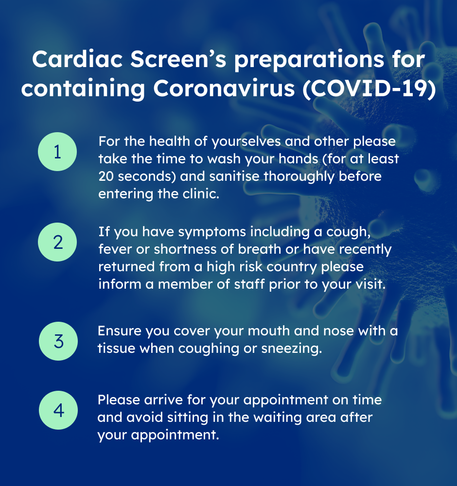
Please click here to view Cardiac Screen's preparations for containing Corona Virus

Balppa House, 57-61 Newington Causeway, London SE1 6BD


 Pay Online
  Bookings


Arterial and Venous Leg
A consultation with a cardiologist
 Make An Enquiry

This test uses sound waves to show an ultrasound picture of the structure of an artery and the amount of blood flowing through it .Gel is applied to the skin, and a probe is placed over an area. A duplex ultrasound helps to tell where in the arteries the narrowing or blockage is and how much it is narrowed. It can be difficult to obtain a picture if you are very overweight or if you have a large build-up of atheroma (fatty material) in your arteries. This test can help to decide which treatment would be most suitable for you.
The most common reason for a venous ultrasound exam is to search for blood clots, especially in the veins of the leg. This condition is often referred to as deep vein thrombosis or DVT. These clots may break off and pass into the lungs, where they can cause a dangerous condition called pulmonary embolism. If the blood clot in the leg is found early enough, treatment can be started to prevent it from passing to the lung. A venous ultrasound study is also performed to: determine the cause of long-standing leg swelling. In people with a common condition called “varicose veins”, the valves that keep blood flowing back to the heart in the right direction may be damaged, and venous ultrasound can help the radiologist decide how best to deal with this condition.
-
Vascular ultrasound imaging is the examination of the body’s circulatory system.
-
It is performed for:
-
DVT Deep vein thrombosis ( blood clot ) in arms or legs
-
Arteries for flow limiting stenosis (usually leg arteries)
-
Peripheral veins to assess varicosities and venous reflux (insufficiency)
-
Thoracic outlet syndrome
-
Swollen legs or arms sometimes caused by a blood clot.
-
Pain in the legs after walking a certain distance is sometimes caused by reduced
-
Arterial blood flow to the legs by disease (intermittent claudication).
-
Varicose veins.
Blood clots may form when something slows or changes the flow of blood in the veins.
Risk factors include:
-
After a pacemaker catheter has been passed through the vein in the groin
-
Bed rest
-
Cigarette smoking
-
Family history of blood clots
-
Fractures in the pelvis or legs
-
Giving birth within the last 6 months
-
Heart failure
-
Obesity
-
Recent surgery (especially hip, knee, or female pelvic surgery)
-
Too many blood cells being made by the bone marrow (polycythemia vera), causing the blood to be thicker and slower than normal
You're also more likely to develop DVT if you have any of the following conditions:
-
Blood that is more likely to clot (hypercoagulability)
-
Cancer
-
Taking estrogens or birth control pills. This risk is even higher if you smoke.
DVTs are most common in adults over age 60, but can occur at any age.
Sitting for long periods when traveling can increase the risk of DVTs. This is most likely when one or more of the risk factors listed above are also present

Pricing
From only £500.00
Arterial Leg Ultrasound Test is Available at a Special Reduced Rate!

Location
Arterial Leg Ultrasound is available at
The Medical Specialist
Balppa House
57-61 Newington Causeway
London
SE1 6BD

Appointments
Same Day Appointments
You can have quick access with same day appointments.
Self Referral
If you are self-referral, our staff would be happy to get a brief history of your symptoms and advise you appropriately with what you will require and which consultant you will need to be booked with.
 Refer Yourself
Contact
Please contact us by clicking the button below, or speak directly to one of our dedicated Patient Care team directly on 020 7403 5294
Cardiac at Home Services
Our unique cardiac at home service means we come to you to perform this test in the comfort of your home.
Do You Qualify?Client Reviews


My appreciation for yet another friendly and professional consultation. Never a welcome undertaking for me but you make it as easy as it can be. Thank you!

Professor Rymer was really helpful and answered all my questions gladly. She really listened and helped me understand her thoughts on my condition. I felt very reassured afterwards!
As usual the reception was courteous and welcoming, emphasised by cups of tea after our 2 hour journey.The Professor's explanation of my condition was clear and very helpful.
Dr. Coutts and his assistant Rekha Dave were both very knowledgable, professional and compassionate. I was very appreciate of Dr. Coutts' assessment of my symptoms and his suggestions for improvem ...ent. Rekha Dave administered the treadmill test and made the process very comfortable for me. I definitely recommend their service to anyone with heart issues.

Price From only £500.00
Arterial Leg Ultrasound
A consultation with a cardiologist
Health Insurance